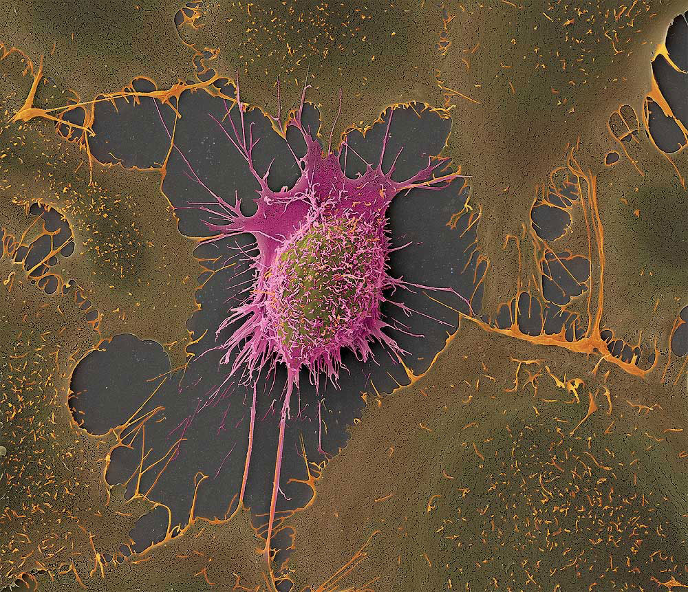
Depicted are cells from a human hepatocellular carcinoma (HepG2). A tumour cell line established from a 15 year old Caucasian boy. These cells have 55 chromosomes instead of 46 as in healthy humans. Cancer cells lose the ability for correct program of dying and consequently proliferate uncontrolled. This fact can be used by researchers not only to study cancer itself, but to use them as a study model, which proliferates indefinitely. In contrast primary established cells from tissue normally die off after a certain number of divisions.